Physical Address
304 North Cardinal St.
Dorchester Center, MA 02124
Physical Address
304 North Cardinal St.
Dorchester Center, MA 02124
So how could this new cell have eluded scientists and doctors for so long? In a way, it didn’t. Plekas and his graduate students have scoured centuries of scientific papers for any lost traces of fatty cartilage. They found a clue in an 1854 German book by Franz Leidig, a contemporary of Charles Darwin. “Anything and everything he could stick under the microscope, he did,” Pleikas said. Leydig’s book described fat-like cells in cartilage samples from rat ears. But 19th-century equipment couldn’t extend beyond that observation and, realizing that a more accurate census of skeletal tissue could be valuable for medicine, Plicus decided to crack the case.
His team began their investigation by looking at the cartilage sandwiched between a thin layer of skin in mouse ears. A green dye that preferentially stains fat molecules reveals a network of squishy blobs. They isolated these lipid-filled cells and analyzed their contents. All your cells have the same library of genes, but those genes aren’t always active. What genes do these cells express? What protein slush around inside? This data revealed that lipochondrocytes actually look molecularly very different from fat cells.
They next questioned how lipochondrocytes behave. Fat cells have one unequivocal function in the body: to store energy. When your body stores energy, cellular stores of lipids swell; When your body burns fat, the cells shrink. Lipochondrocytes, it turns out, don’t do anything like that. Researchers studied the ears of rats placed on a high-fat versus calorie-restricted diet. Despite rapid weight gain or loss, ear lipochondrocytes did not change.
“This immediately suggested that they must have a completely different role that has nothing to do with metabolism,” Pleikas said. “It has to be structured.”
Lipochondrocytes are like balloons filled with vegetable oil. They are soft and amorphous but still resist shrinkage. It contributes meaningfully to the structural properties of cartilage. Based on data from mice, the tensile strength, elasticity, and stiffness of cartilage increased by 77 to 360 percent when comparing cartilage tissue with and without lipochondrocytes—suggesting that these cells make cartilage more flexible.
And structural gifts seem to benefit all kinds of species. In the outer ear of Pallas’ long-tongued bats, for example, lipocartilage lies beneath a series of ruffles that scientists believe tune them to specific wavelengths of sound.
The team also discovered lipochondrocytes in human fetal cartilage. And Lee says the discovery ultimately explains something that reconstructive surgeons often see: “Cartilage always has some slippage to it,” he says, especially in young children. “You can feel it, you can see it. It is very clear.”
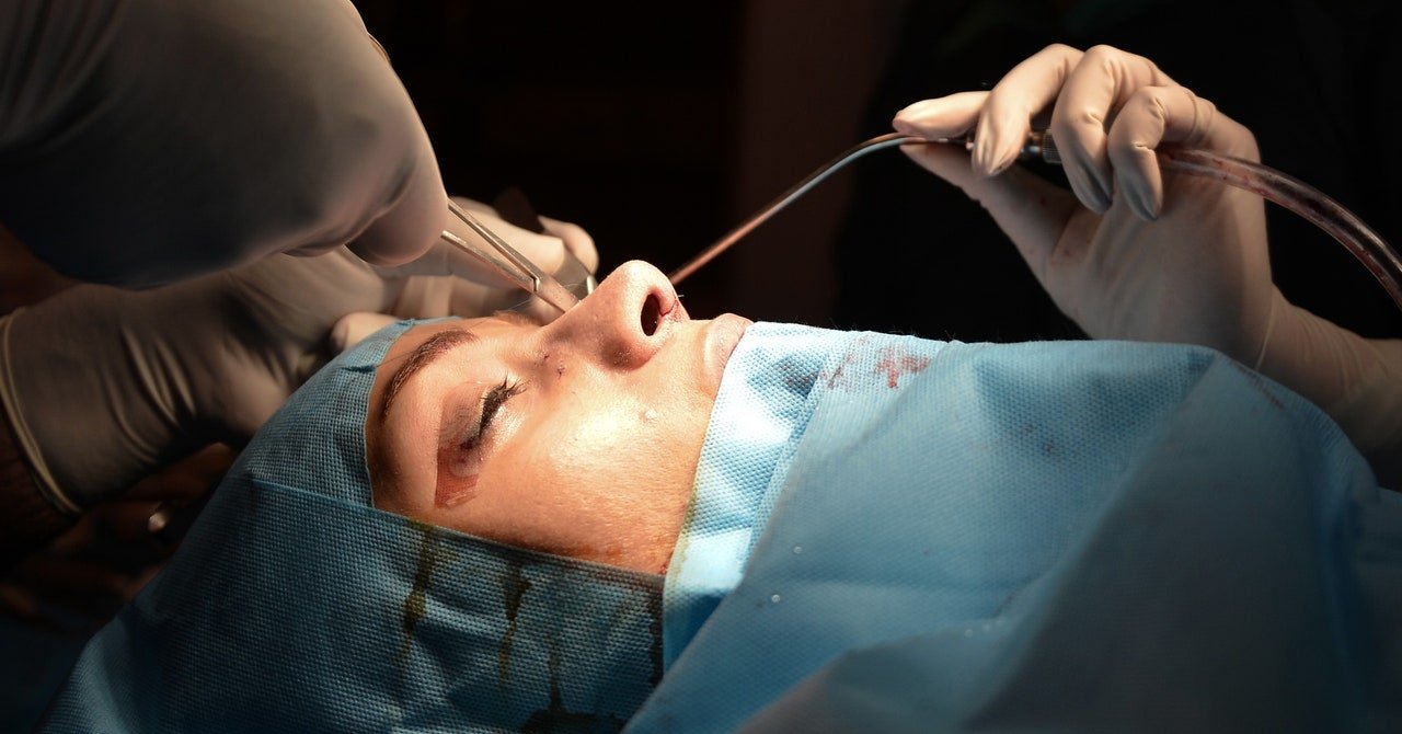
New findings suggest that lipochondrocytes fine-tune the biomechanics of some of our cartilage. A stiff fold of cartilage protein without lipids is more durable and is used to make weight-bearing joints in your neck, back, and—yes, you got it—ribs, a traditional source of cartilage for implants. “But when it comes to more complex things that are actually flexible, bouncy, elastic — the ears, the tip of the nose, the larynx,” says Plekas, that’s where lipocartilage shines.
As for the procedures involved in modifying these body parts, Plekas envisions one day growing lipocartilage organoids in a dish and 3D-printing them into any desired shape. Lee, however, cautions: “Despite 30 or 40 years of study, we’re not very good at making complex tissues,” he says.
Although such an operation is far off, studies suggest that it is possible to grow lipochondrocytes from embryonic stem cells and safely isolate them for transplantation. Lee figures that regulators won’t greenlight using embryonic cells to grow tissue for non-life-threatening conditions, but said he would be more optimistic if researchers could grow transplantable tissue from adult cells from a patient. (Plikas says a new patent application he filed covers using stem cells from adult tissue.)
Lipochondrocytes update our understanding of what cartilage should look and feel like—and why. “When we try to create, say, the nose, sometimes we can use [lipid-filled cells] for a little padding,” Lee says. Lipocartilage could one day fill that void as a growable, transplantable tissue—or it could inspire better biomimicry materials. “It could be both,” he says. “It’s exciting to think about. Maybe it’s That’s one thing we’ve lost.”